
Bone Biology
Our bone cell biology program focuses on three core aspects of musculoskeletal science: the fundamental biology of osteoblasts and osteoclasts in health and disease; the effects of ionising radiation on skeletal tissues; and the interactions between tumour cells and the skeleton in orthopaedic cancers.
We are looking at the fundamental ways that tumours spread around the body, and working with Mesenchymal Stem Cells derived from the Periosteum and Bone Marrow to look into these cells ability to regenerate bone fractures.
In collaboration with the Queen's Veterinary School Hospital and within the Department of Veterinary Medicine at the University of Cambridge we can also share the expertise and access to resources in the Surgical Discovery Centre, Clinical Pathology, Histology and more.
Projects
Innovative tissue engineering approaches for bone regeneration, which involves in vitro and in vivo studies on mesenchymal stem cells in combination with different 3D printed scaffolds.
Effects of bortezomib on the radiation sensitivity of Ewing's sarcoma
Working with different cell lines to investigate the mechanisms involved in osteosarcoma cell growth
Exploring the regenerative capacities of Mesenchymal Stem Cells, comparing their source of origin (Periosteum or Bone Marrow), with in vitro work (culturing and expanding cell populations), checking for osteogenic markers, cell characterization, differentiation and functionality under certain conditions.
Radiobiology of bone
Investigating Mesenchymal Stem Cells: Processing and Imaging Techniques

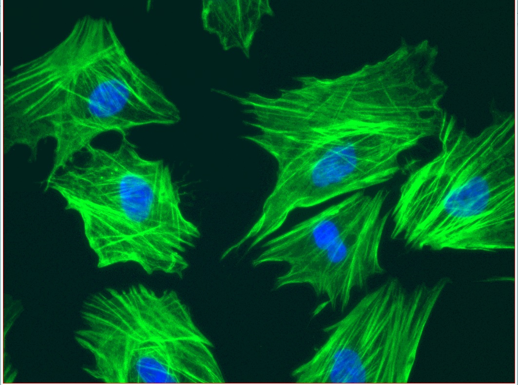
Mesenchymal stem cells (MSCs) Immunofluorescent staining and imaging using Alexa Fluor® 488 (green) labelled beta-actin antibody + DAPI (blue) for nucleus staining .
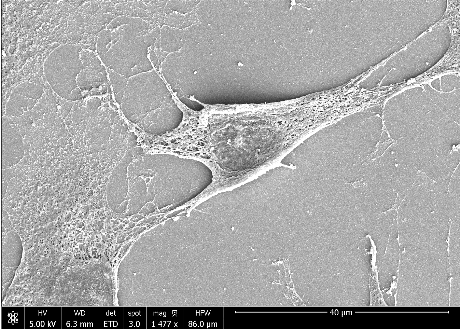
Scanning electron microscopy (SEM) images of mesenchymal stem cells (MSCs)


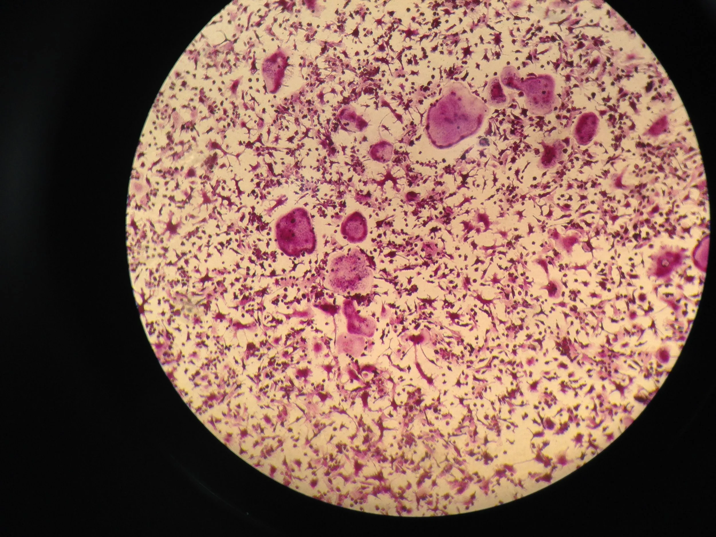
Inverted light microscope x 20 image showing Haematoxylin stained MSCs
Periosteum seen under the scanning electron microscope
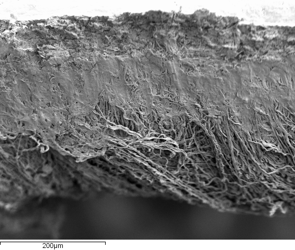

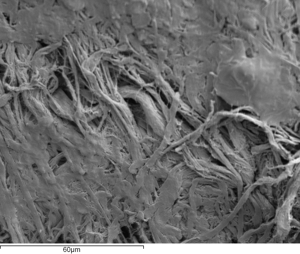
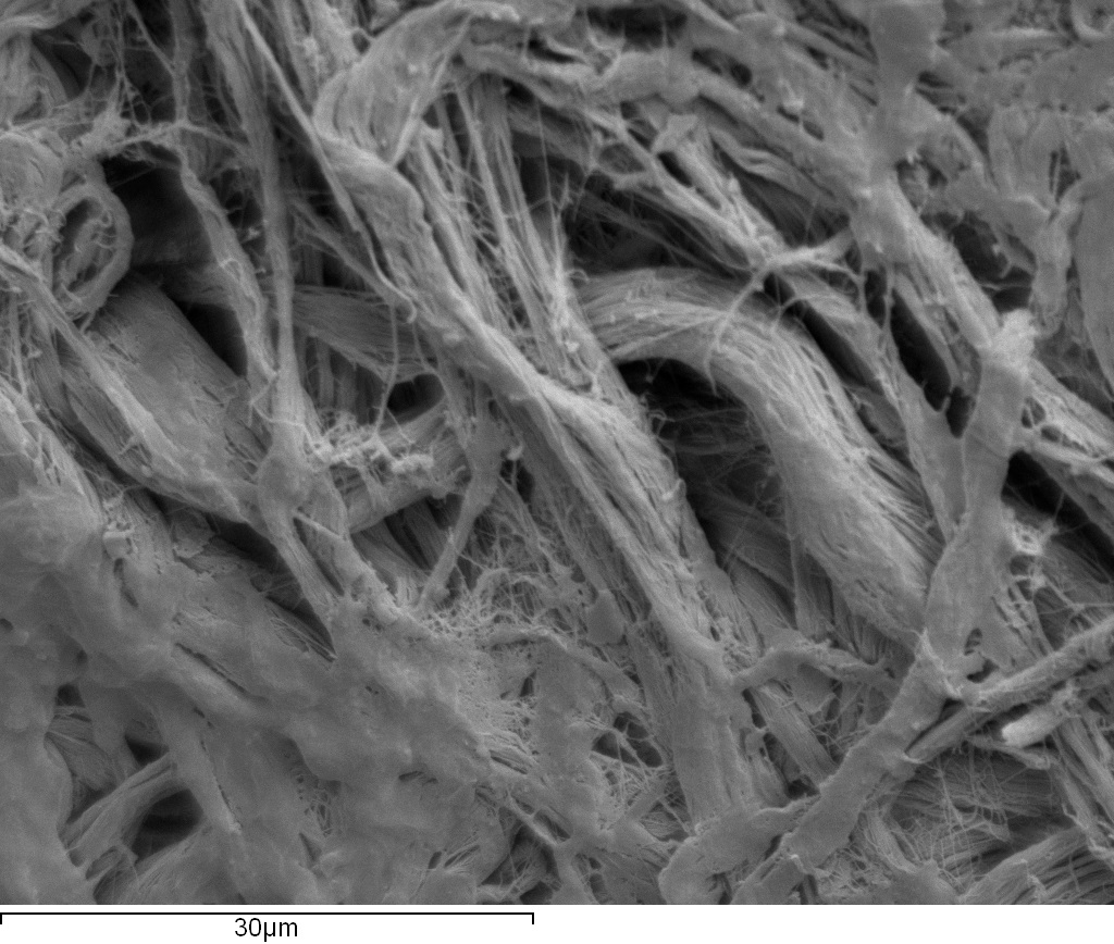
Periosteum is a thin vascular membrane that covers the external surface of bone except for the articular surface of long bones (Roberts et al., 2015). It consists of two layers: an outer fibrous layer containing fibroblasts dispersed in between collagen fibres, and an inner cambium layer which contains skeletal progenitor cells and osteoblasts (Allen et al., 2004). The periosteum serves as an attachment site for tendons, ligaments and muscles (Chanavaz et al., 1995). It plays an important role in bone fracture healing, when periosteal progenitor cells undergo expansion, followed by differentiation into osteoblasts and chondrocytes (Colnot et al., 2009; Ozaki et al., 2000). This process forms a callus which is later replaced by new bone and eventual remodelling to restore the original shape of the bone (Colnot et al., 2009; Ozaki et al., 2000).
Our Equipment
Evos XL Core Imaging System
We use the Evos XL Core imaging system in our Cell Culture Lab for viewing routine cell and tissue culture. Replacing the eyepieces of a standard Microscope with a high-resolution monitor allows for multiple users to work together, enabling collaborations and enhancing teaching capabilities.
The microscope takes up a fragment of the footprint of other microscopes, and is lightweight, offering great flexibility and ease of use.
We can also store some great images on USB, for teaching and review.
Biodent indentation platform
The BioDent is a Reference Point Indenter that allows for direct measurement of bone tissue strength. The reference point shields the inner probe from soft tissue, and allows for a reference for displacment measurements at the site of contact.
Makerbot Replicator 2x
We use the Makerbot to print 3D bones and cutting guides in preparation for our surgeries. using CT scans we can reconstruct the bone and use it to help design and prepare surgical tools.


